Sunday, November 22, 2009

Harnessing the Power of Stem Cells

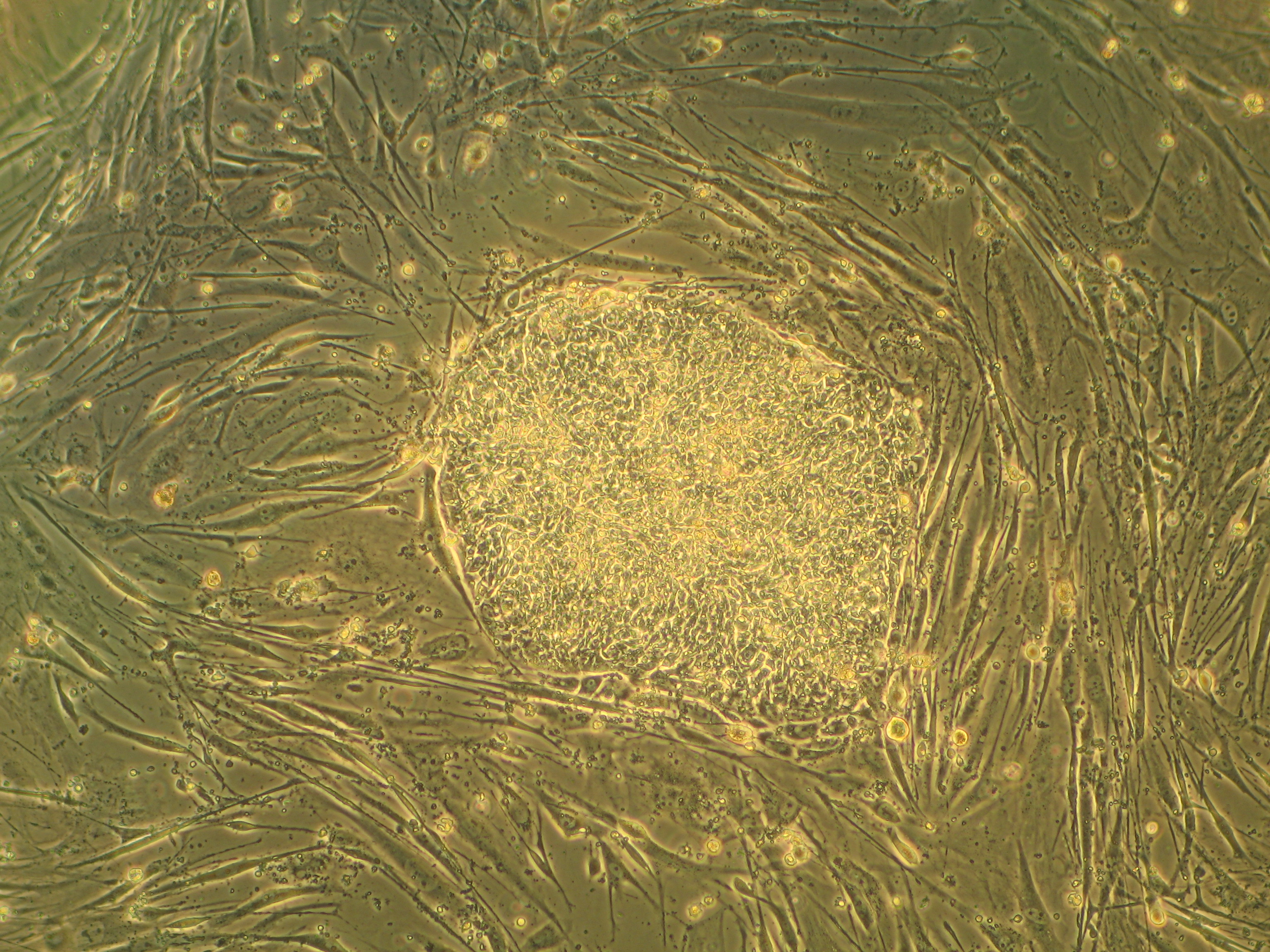
Researchers at the Wellcome Trust Center for Stem Cell Research have recently been able to uncover a potential crucial link that helps to uncover the remarkable properties of stem cells. This research team at the University of Cambridge has been able to find the last and most crucial step in a complex procedure that results in stem cells having their ability of being able to develop into different cells within the body such as skin cells or even liver cells. The reports which were published in the journal Cell, have resulted in greater efforts for the harnessing of stem cells to be able to treat any medical conditions.

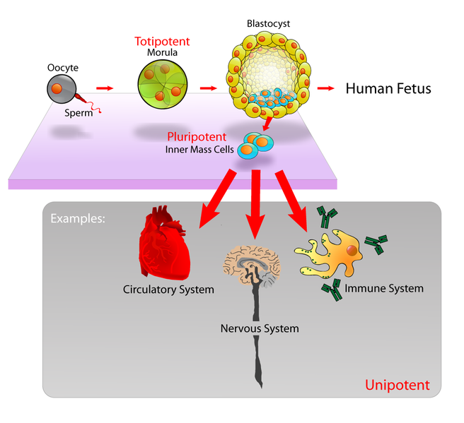
There has been a lot of research that has been conducted in the field of stem cells during the last few years. It is now possible for scientists to transform adult brain or skin cells into embryonic stem cells. Very similar to natural stem cells, such embryonic stem cells can be adapted to function just like any other cells of the body. This ability which is known as pluripotency, is now being used as the basis for the development and modification of stem cells that will one day be able to help in fighting diseases such as Parkinson’s, Alzheimer's or even diabetes.

Dr Jose Silva along with his colleague Dr Jennifer Nichols of the Cambridge research team state, “Inspite of having uncovered many exciting developments, we were still a long way away from actually finding out how cells become pluripotent. It was a mystery that had to be solved in order for us to be able to create safe, reliable and efficient methods for the generation of these medical cells. It is vital that we are able to understand how and what exactly brings about this process.”

Funded by charitable institutions, it was discovered that a protein called Nanog helps in achieving pluripotency. “We always knew that Nanog was a very important substance, although we did not know how exactly it helped. We now know that it is this protein that aids in a complex process helping in pluripotency." If there were no Nanog, it would be impossible for the embryo to develop or to be reprogrammed into adult cells. The next step in the research process is to actually find out how Nanog can help in influencing other molecules that it is surrounded by."


This research conducted was supported by the Wellcome Trust, the EC Framework and the Biotechnology and Biological Sciences Research Council.


Wednesday, November 18, 2009

Monkey Teeth Help in the Stimulation of Brain Cells

A newly conducted study by researchers at the Yerkes National Primate Research Center, Emory University, has shown that stem cells in teeth can be used to aid in the generation and growth of different neural cells. This study which was recently made available in ‘Stem Cells’ October issue details how it is possible for dental pulp stem cells to actually bring about regeneration and aid in cell therapy thereby helping any individual who is undergoing therapy in relation to the central nervous system.


Stem cell research is usually divided into two main branches out of which dental stem cells is one. These dental stem cells which are adult stem cells have the ability of regeneration into many different types of cells thereby increasing the possibility of therapeutic treatment for potentially dangerous diseases such as Parkinson’s and Huntington’s. Earlier tests have already concluded the ability of dental pulp stem cells of being able to aid in the re-growth and restoration of craniofacial and dental cells.

The team of scientists at the Yerkes researcher at Emory University, led by Anthony Chan, DVM, PhD, conducted their experiments by using dental pulp stem cells extracted from the teeth of the rhesus monkey better known as the rhesus macaque, and then directly implanting them into the hippocampus of mice. “We noted that there was greater formation of neurons and at the same time an increase in the stimulation of growth of new neural cells. Our research has shown that it is possible to stimulate the growth of neurons through dental pulp stem cells. We now know that this will result in great therapeutic benefits and that dental pulp stem cells will soon one day be able to achieve a much broader goal,” says Chan, an assistant professor at the Emory School of Medicine studying human genetics.

The fact that it is possible to isolate dental pulp stem cells at any age from any patient by a simple visit to the dentist interests Chan as it is an indication of the possibility of banking such dental stem cell. “The chances of rejection by the body would greatly be reduced if it were possible to use one’s own stem cells for therapy,” says Chan.

The next step in the experiment for Chan and his research team is to determine whether dental pulp stem cells from Huntington suffering monkeys would help to improve, augment as well as enhance the development of any brain cells in mice.

Monday, October 19, 2009

Parkinson’s Disease Can Soon be Cured

A particular antibiotic mutated by researchers at the University of Florida has represented the first step in the curing of Parkinson’s disease.  An article that was published in the September issue of Molecular Therapy has shown that a particular antibiotic that was mainly used to try and cure Parkinson’s disease can also function as a cure for gene therapy. The study which was conducted in rats now shows how therapeutic genes which when delivered to the human brain can help in the treatment of Parkinson's disease.


adeno associated virus (source Wikipedia)

Says Ronald Mandel, a professor of neuroscience at the Powell Gene Therapy Center and University of Florida‘s McKnight Brain Institute, “Most prior experiments that made use of any growth factors that grew naturally in the body to divide and grow cells as a means of rejuvenating dying brain cells failed due to the fact that they were injected too late. However, newer findings suggest that it is possible for the brain to produce a neurotransmitter known as dopamine mainly thanks to gene therapy.” Patients in Parkinson’s disease often face a shortfall in the production of dopamine, but now due to the latest findings of the research, there would be an increased likelihood of success. Dopamine is very essential as it helps in the communication and coordination between cells.

“We have spent over 10 years trying to design a gene delivery vector that can help in safely transferring the genes required for Parkinson’s. We now believe that it is possible to intervene at the earliest stages of the disease as some of our earlier interventions failed to save dopamine-producing connections in patients mainly due to the fact that they were given to suffering patients only during the latter stages of the disease and as such there were very few dopamine-producing connections left,” says Mandel.

The Mandel led team of scientists used an adeno-associated virus that was capable of inducing dopamine cells into animal brain cells which could then result in the production of GDNF. This GDNF is vital as it helps the dopamine-producing neurons survive during the process of development of the brain of adults.

The virus was engineered by scientists with two genes so as to be able to act in connotation with each other to produce protein. By using an antibiotic called dietary doxycycline, the progression of the genes could be slowed down. The amount of the antibiotic determines the regulation of production of the protein. In other words, it is even possible to reduce this protein which in turn can give medical investigators the option of regulating gene therapy after delivery of the treatment.

Monday, October 5, 2009

Making Use of Stem Cells as a Means of Treating Male Infertility

A recently conducted survey has revealed that it might be possible to treat male infertility through the use of bone marrow stem cells. The research conducted, which was published in an issue of ‘The American Journal of Pathology’, showed that when stem cells of bone marrow were transplanted into the testis of a male, there was the potential for overcoming testicular failure.

Under normal circumstances, when a couple tries to conceive a child, but is unable to do so, it is more often than not the male who is responsible. Due to differentiation and proliferation of the germ cells or even due to any type of dysfunction from supporting cells, a male might experience infertility. The research study which was carried out under the direction of Dr. Ronald S. Swerdloff, was undertaken with the intention of replacing nonfunctioning germ or other cells with stem cells.


At the Harbor-UCLA Medical Center, mice which expressed the green fluorescent protein (GFP) were used for the collection of bone marrow stem cells. Using chemically modified mice so as to induce infertility, these green cells were then injected into the testes of the mice and the results were noted. For the entire 12 week period, the GFP cells that were injected into the mice managed to survive and took up residency in the testes. These cells managed to mirror the characteristics of germ cells, an indication of the separation of such cells. The research also found that many of the differentiated cells were located very close to the native cells perhaps due to the influence of the local cellular environment.


GFP 3D structure and its extraction source - jellyfish Aequrea Victoria (source Wikipedia)

By verifying certain proteins on the surface of the donor cells, Dr Ronald S. Swerdloff and his team found that the germ and supporting cells expressed types of proteins that were usually found only in differentiated cells. Says Dr. Swerdloff, “Our findings indicate that it is possible for bone marrow stem cells to multiply and act like the germ or supporting cells that are responsible for sperm production in the testes. However, additional factors might be involved as can be seen from the fact that the germ cells did not completely separate into sperm.”


There is the need for further studies that can list the various hormones that are required for the production of sperm in the transplanted model. Further testing can also reveal which are the specific stem cells that can differentiate and colonize in the testes. These studies could have a great implication in being able to treat testosterone deficiency or male inefficiency.

Thursday, September 24, 2009

Developing Sperm Used in the Creation of Stem Cells


Scientists at Johns Hopkins University School of Medicine have realized that the discovery of how to revert adult cells into their elemental stem cell state might hold the key to what exactly stem cell therapy can accomplish. Through their research, these scientists have been able to discover those key molecular players that have mainly been accountable for the fruit fly sperm cells reversion process. In an online report that was published in Cell Stem Cell, it was noted that two very important proteins- Jak and STAT- had been responsible for redirecting sperm becoming cells into stem cells. An associate professor of cell biology at the Johns Hopkins University School of Medicine, Dr. Erika Matunis stated that earlier research conducted by the group showed that it was possible to convert sperm into stem cell, although the way to do so still remained a mystery. “Dedifferentiation is an extremely motivating and appealing wonder that has had its occurrence in a variety of stem cell populations and as such, we wanted to know everything that we could about the process,” says Dr. Matunis.


Similar to stem cells, the fly testis also contained nine stem cells that were divided to create daughter cells. Out of these two, one was differentiated into an adult cell or a sperm cell while the other remained a stem cell. The research team at Johns Hopkins University School of Medicine genetically altered the flies in order to find out the reason behind the dedifferentiation of these sperm cells. The flies were altered so that both the cells would transform into sperm resulting in the stem cell population of the testes becoming nothing. A week after the genetic modification, it was found that the two stem cells had repopulated from the fly testes.


It had long been suspected that two proteins, Jak and STAT were not only known to work together in helping stem cells, but were also responsible for dedifferentiation. “By genetically altering the flies, we were able to reduce the amount of activity in the STAT and Jak proteins. We now know that it is possible to interfere with the process of dedifferentiation by interfering with these two proteins in the fly testis.


The team realized that fewer cells would revert back to stem cells if there was interference with the two proteins. By counting the number of cells it was found that unlike normal Jak-STAT activity where 97% of the testes regained stem cells, such interference caused merely 60% of the testes to regain stem cells.


Wednesday, September 16, 2009

Human Stem Cells Used to Manufacture Red Blood Cells that can Glow

 Scientists at the Monash University in Victoria have managed to modify human embryonic stem cells so that they can ‘glow’ when transformed into red blood cells. These customized embryonic cells are representations of the first focal step taken towards the generation of completely functional red blood cells. Although human embryonic stem cells have the capability of being modified to fit any required cell within the body, it still remains a challenge to try and turn these embryonic cells in any specific helpful cells. For this reason, the findings at Monash have represented a major breakthrough in scientific study.


Studies are now being conducted on such human embryonic stem cells so as to try and find an answer to a variety of complex questions regarding human development. One of the primary objectives in the study of such stem cells is to find out how they can become differentiated cells forming part of the organs and tissues of the body. Many diseases such as cancer as well as birth defects have been attributed to the abnormal separation of such embryonic cells. To completely understand the controls of such cells, human embryonic stem cell research is currently being carried on.


Led by Professor Ed Stanley and Professor Andrew Elefanty as well as other leading scientists from the Murdoch Children’s Research Institute, the findings of the research which were funded by the Australian Stem Cell Center (ASCC) were published in the journal, ‘Nature Methods’. The ASCC helps to track the demarcation and transfiguration of the embryonic stem cells into red blood cells.
The ASCC’s Scientific Director, Professor Joe Sambrook was quoted as saying, “The fantastic work ethics of the Elefanty-Stanley team has finally paid dividends and has brought to light the differentiation pathway which has finally led to our better understanding of the manifestation of adult hemoglobin genes.”
For quite some time, it had remained a scientific challenge to be able to turn human embryonic cells into other types of cells. Through their research, the team managed to incandesce red into the ErythRED embryonic stem cells when mixed with hemoglobin. This brings hope to the fact that in future, researchers would be able to enhance the conditions that bring about the thriving of such cells. This ErythRED cell line can lead to the improved creation of red blood cells and can also be used as a means for monitoring any transplanted animal model cells.
The research was supported by the Juvenile Diabetes Research Foundation, the National Health and Medical Research Foundation and the Australian Stem Cell Centre.

Saturday, September 12, 2009

Stopping cancer stem cells through lung cancer Oncogene

At the Mayo Clinic in Florida, scientists have unearthed that  PKCiota – a lung cancer oncogene- has been responsible for the proliferation of lung cancer stem cells. These powerful and very rare stem cells are responsible for the manufacture other cells that lead to the building up of various tumors. These cancer causing stem cells are also very resistant to chemotherapy.

PKCiota is a human oncogene, in other words, it is an abnormal gene that is used by various malignant cancers to grow and survive. Most lung cancers are able to genetically alter as well as over-express this PKCiota which in turn results in the patient becoming very weak and having a very low rate of survival.
In an issue of Cancer Research, a study showed that it was capable of stopping the growth of any cancerous stem cells through the use of a particular agent called aurothiomalate. This aurothiomalate is currently being tested at the Mayo Clinic and is in a clinical trial of phase I.
Alan Fields, chair of the Department of Cancer Biology at Mayo Clinic and professor of pharmacology in the College of medicine, who is also the senior analyst in the study said, “Our research has proved that PKCiota is needed at the earliest stages and aids in the growth tumor initiating cancerous stem cells which ultimately leads to lung cancer.”
“Most common lung cancers are caused by these lung cancer stem cells and so, it is vital that such cancerous stem cells be disrupted in order for any therapeutic treatment to work. Our impending research has shown that aurothiomalate can in fact be used to effectively target such stem cells.”
Used once upon a time to treat rheumatoid arthritis, aurothiomalate has now also been proved to be very effective in targeting PKCiota. There is a trial phase I that is currently being conducted on various patients at the Mayo Clinic in Arizona and Minnesota and depending upon the results in phase I, phase II has been scheduled to combine aurothiomalate with various other agents that are capable of slowing down the growth of such cancerous cells.
Although, it was previously known that PKCiota was able to maintain tumor growth, these new findings show that PKCiota is also responsible for the initial development of lung cancer. Scientist for long have known that cancer stem cells were capable of initially bringing about tumors, however, the discovery of aurothiomalate has now proved that these stem cells are not as resistant to tumors as previously thought.

Monday, September 7, 2009

Human cell transplantation is able to prolong survival of mice

A recently conducted study has shown that the successful transplantation of human neural stem cells has been able to provide substantial improvement in a rare hereditary neurodegenerative disease. These findings which were published in the September issue of Cell Stem Cell show that a critical enzyme which was missing from the brains of the experimented mice was provided for by the transplanted cells. This study has proved that there might be hope for those patients that are currently suffering from any types of devastating or untreatable diseases.

One type of neurodegenerative disease that is found in children is the Infantile Neuronal Ceroif Lipofuscinosis (INCL). Also known as Batten disease, INCL is formed by a deficiency of the enzyme palmitoyl protein thioesterase-1 (PPT-1) that is created by a particular gene. A deficiency of PPT1 results in an accumulation of a cellular lipid known as lipofuscin. Any accumulation of this lipofuscin can lead to visual impairment, reduced motor and cognitive skills, neuron death, seizures and also cause premature death.

Currently, it is almost impossible to get PPT1 enzymes in the brain and so intravenous enzyme treatment is not an apt solution. However, it has been conjectured that donor cells that have been transplanted would be capable of supplying the needed enzymes directly into the brain. A mouse model that is capable of exactly replicating many aspects of human disease has been created and is now being used to find whether such stem cell replacement would be beneficial or not. Led by Dr. Nobuko Uchida, a study was carried out to test this hypothesis. “We made use of nontumorigenic, normal and nongenetically modified human cells to ably deliver the enzymes into the modeled mice. We transmitted these neural stem cells as they are self renewing and provide a permanent production of any missing enzymes.” The transplanted human neural stem cells were capable of matching and delivering the required enzymes to the modeled mice, while at the same time also providing a production of PPT1 to bring about significant improvement. After the replacement therapy, the INCL infected mice showed signs of reduced loss of motor coordination, less lipofuscin and greater neuroprotection.

These findings have shown that the direct transplant of such stem cells into the brains of those that suffer from INCL might be able to provide a long lasting and continuous supply of the missing PPT1 as well as some therapeutic benefits, thereby providing a potential medical breakthrough in combating such a disease.

Tuesday, September 1, 2009

Gene therapy not the cause of arthritis patient death

Medpage Today published on July 08, 2009 reported a 32 year old women death who participated in the gene trial therapy for rheumatic arthritis. The patient died after 22 days of treatment. This incident has put a halt to all the clinical tests that was going on related to gene therapy. Actually the researchers were testing a drug named experimentally as tgACC94. This drug is based on the recombinant adeno-associated virus (AAV) derived vector. It carries the tumor necrosis factor receptor (TNF-receptor) gene to block the inflammation.

After investigation the researchers say that the death of the patient is not because of the gene therapy treatment for rheumatic arthritis. It is because of the wide spread histoplasmosis. Histoplasmosis is a normal fungal infection but in this case it has gone deadly as the result of immunosuppressant therapy. The patient was under immunosuppressant therapy for few years she was under the medication of adaimumab, methotrexate and prednisone. These drugs have only increased the risk of histoplasma fungus, said the researchers. They also add that the combination of histoplasmosis and retroperitoneal hematoma has only led to patients death. It is a well known thing that the risk is more when a patient affected by histoplasmosis takes TNF antagonists. The important explanation the researchers say is that she was already infected by the histoplasmosis fungus when she was receiving her second dosage of tgAAC94 as a part of her gene therapy. After the second dosage she was suffering with large number of symptoms like fever, vomiting, abdominal pain etc. The symptoms started getting worse day by day and she was hospitalized. Finally she was suffering from large abdominal hematoma, liver damage and kidney failure. After three weeks of suffering from all these symptoms from the day the second dosage for gene therapy was given she died on 24th July. Some people say, the death can be avoided if the researchers had taken enough care of the patient at the beginning itself when she started showing some negative symptoms. If she would have brought under complete monitoring at that time itself definitely her death would have been avoided.

Elizabeth L.Hohman, MD, of partners human research committee in Boston, wrote in an editorial that this incident has taught a number of lessons to the researchers, mainly making them to concentrate about the endemic infections in the area where the patients are residing. This incident has cautioned all the researchers around the globe. This has made them to take a keen note of all the endemic diseases that are in the prevailing in patient areas who all are receiving TNF antagonist drugs. And also in addition to that the researchers have come to know about the importance of a proper complete monitoring system of the patients who fall ill when they are under such researchers. Because of this negative result in gene therapy people will come to a thought that acceptance of a gene therapy approach for non genetic diseases such as arthritis is marginal and the serious adverse effect of the treatment could even destroy the entire activity.

Sunday, August 30, 2009

Nanoparticles combined with 'suicide' genes slowed ovarian tumor growth

It is very good to hear that nanoparticles have slowed ovarian tumor growth in mouse experiments since it might help one day many women suffering from late stage ovarian cancer. Ovarian tumor growth was a big issue for the past years though the ovarian tumor at initial stages was treated surgically followed by chemotherapy the treatment for ovarian cancer in advanced stages had no effective results. It is disproportionately deadly because it lacks any clear early detection or screening, meaning they are diagnosed only after they have reached advanced stages. Over 75% of ovarian cancers are diagnosed at an advanced stage. Currently the treatment for ovarian cancer is give by chemotherapy but in many cases the cancer returns back and there are no good therapies for recurring and advanced stage ovarian tumors. This new treatment reported by July 31 issue science daily, delivers a gene that produces the diphtheria toxin, which kills cells by disrupting their ability to manufacture proteins. This toxin is normally produced by cornynebacterium diphtheriae. The other good thing about this treatment on advanced ovarian tumor growth unlike chemotherapy which acts on both good and bad cells is that it acts only on the infected tumor cells leaving back the good ones said the lead researcher Janet Sawicki, Ph.D., a professor at the Lankenau institute for medical research. It is also said that Sawicki along with her team experimented the efficiency of cationic biodegradable beta-amino ester polymer as a vector for the nanoparticle delivery of a DNA encoding diphtheria toxin suicide gene. They are called poly beta amino esters because they are the new nanoparticles and are made with positively charged, biodegradable polymers known as poly beta amino esters. When mixed together, these polymers can spontaneously assemble with DNA to form nanoparticles. The polymer-DNA nanoparticle can deliver functional DNA when injected into or near the targeted tissue. These were injected into mice with ovarian tumors. The result was not happy so the team again did the management of nanoparticles in three different mouse models. This showed a very good suppression of tumor size with minimal non-specific cytotoxcity. It did not have toxic effects of chemotherapy because the gene is engineered to be over expressed in ovarian cells but is inactive in other cell types.

mucinous ovarian tumour (source: wikipedia)

The main thing about treating this advanced stage ovarian cancer is that finding the target where these cancerous cell prevail was very difficult said Edward Sausville, M.D., Ph.D., an associate editor of cancer research and associate director for clinical research at the Greenebaum Cancer Center at the University of Maryland who is working in the Oncology department studying ways to kill tumors for a long time. But now Sausville is happy that this new method of treating ovarian cancer can find the target in many different ways.

In count to ovarian cancer, these nanoparticles have demonstrated potential for treatment of a variety of diseases, including prostrate cancer and viral infection. In future studies the team has plans to examine the effectiveness of nanoparticle-delivered diphtheria genes in other types of cancer, including brain, lung and liver cancers.

Saturday, August 29, 2009

Adult stem cells

Adult stem cells are stem cells that are found in the tissues of organ in developed human beings. These cells are deep hidden within organs and are surrounded by millions of cells. The adult stem cells help in development and regeneration of the organ in which they are found. The term adult is used to indicate that these stem cells are further along the path of differentiation than are embryonic stem cells. Scientists use the term somatic stem cell instead of adult stem cell where somatic refers to cells of body.

bone marrow mesenchymal stem cells

Bone marrow mesenchymal stem cells

The adult stem cells are mutipotent, meaning they are able to produce a limited range of specialized cells, for example the various kinds of blood cells, bone cells, or muscle cells. The adult haematopoietic and the blood forming stem cells that are found in bone marrow are used in various transplants for 40 years. There are two types of stem cells that were found nearly fifty years ago in the bone marrow they are haematopoietic stem cell and mesenchymal stem cell. The haematopoietic stem cell helps is the formation of blood cells of the human body system and the mesenchymal also know as the bone marrow stromal stem cell make up a small proportion of the stromal cell population in the bone marrow and they can generate bone, cartilage, cells that support blood formation and connective tissue .Now several stem cells are found in human system namely dental pulp derived stem cells, haematopoietic stem cells, mammary stem cells ,mesenchymal stem cells, endothelial stem cells, neural stem cells, olfactory stem cells and testicular stem cells.

From 1960’s to 1990’s many scientists believed that adult brain does not have stem cells but it was said wrong .in later 1990’s scientists agreed that the brain contains stem cells and also the three major types of brain cells namely astrocytes, oligodendrocytes and neurons can be generated from these stem cells.

These adult stem cells are thought to reside in a specific area in the tissue called the stem cell niche. The adult stem cells are very difficult to maintain and grow in culture. Once the adult stem cells are taken out of the human body the speed of multiplying and growing of these cells gets reduced. Scientists are on research to find ways by which they can grow and multiply these cells so that it can help us in injuries or diseases.

The adult stem cell has a wide advantage in therapeutic applications of stem cells. That is as with the help of stem cells several organs of the same type can be replicated, these organs can be used in the testing of various medicines this helps the pharmaceuticals to know the side effects and characteristics of the medicines. The attributes of adult stem cells are demonstrated success in some treatments and stem cells may be genetically matched to patient. The limitations of adult stem cells are they produce limited number of cell types, they are not found in all tissues and difficult to identify, isolate, maintain and grow in the laboratory.

Adult stem cells are very success in curing leukemia and bone marrow transplants. It is so evident that in future with the help of this stem cell we can cure all diseases that are said to be incurable now.

Friday, August 28, 2009

Gene Therapy, new hope for Patients with Hereditary Lung Disease

Science daily reported on august 11th 2009 stated that the Researchers at the University of Massachusetts Medical School and the University of Florida in Gainesville has done wonderful job in the gene therapy trial of boosting protective protein in patients with hereditary lung disease. They have proved to be successful by giving new functional genes to patients who have this hereditary gene problem and the results are positive. This is very good news for people suffering from hereditary lung disease and also this news will bring a good hope in those people. The most interesting news about this therapy is that with just one series of injections our bodies will be able to produce the alpha-1 protein we need till our life time unlike the current existing therapies. The main reason for hereditary lung disease is due to the deficiency of alpha-1 antitrypsin. The people suffering from deficiency of alpha-1 antitrypsin are easy to get affected by any allergies from air as they are not able to produce protein alpha-1 antitrypsin which is normally produced by the liver to protect the various infections that affect the lungs.


The clinical test was practiced in few patients who were affected by this deficiency. An injection of alpha-1protein was given to them in their deltoid muscle in their upper arm. Till one year they were able to produce noticeable amount of alpha-1 antitrypsin. Mark L. Brantley, MD, at UF's College of Medicine he is the professor of medicine, molecular genetics and microbiology in addition to that he is the first author of the study who said the deltoid muscle where the injection is given acts as a factory for making the protein that these individuals are missing. Later as a part of their research they divided nine patients into three groups and were given the adeno associated virus which helps in production of alpha-1 protein. All nine Patients were given injections in their upper arms deltoid muscle. Nine injections were given to them .in each group dosage given was varying. These tests took place at the General Clinical Research Center at Shands at UF Medical Center. After one year the replaced gene was doing its work in three patients successfully who were given the maximum dosage.


The researchers were very happy as there was no rejection of the transferred gene in the patient’s body or the protein that is created newly, though the patients showed some high immune response therapy. That is very good development in gene therapy said Brantley. In the UF Genetics Institute he is a member of the Powell Gene Therapy Center and also he is the Alpha-1 Foundation Research Professor at UF and is a consultant for the organization. Walsh said “alpha-1 community is incredibly grateful for the progress that these dedicated investigators have made” Walsh. The Alpha-1 Foundation’s president and chief executive officer is Mr. Walsh and also he has been constantly supporting various researchers and investigations in gene therapy field for many years. Though currently there is some effective injection of alpha-1 protein derived from human plasma for serious breathing symptoms. They don’t really cure the disease they only slow its progression. So the American Lung Association says that the patients should continue the injections throughout their life. So definitely this research is a boon o many people suffering from serious lungs related problems.

Thursday, August 13, 2009

Embryonic stem cells

Embryonic stem cells are cells that are derived from embryos. They have the potential to get transformed to any desired organ.


In human beings when the fertilization is successful between an egg and a sperm a fertilized egg called zygote is formed. This first divides into two then 4 and so on. Now the embryonic cells are totipotent which means it is capable of giving rise to an entire organism itself. They are also capable of forming extra embryonic supporting tissues like placenta. After five to seven days a multicellular ball of cells called a blastocyst is formed. This blastocyst consists of mass of undifferentiated cells inside it. It looks like a hollow ball made up of two types of cells namely the outer layer of cells called tropoblast which forms the placenta and the inner cluster of cells know as inner cell mass which forms the embryo. This inner cell mass consists of embryonic stem cells. These embryonic cells are no longer totipotent but still pluripotent meaning they are capable of forming all the organs which comprises a human being. But they are not capable of forming placenta or fetus.


There are two key features of embryonic stem cells: pluripotency and the ability to self renew while retaining their undifferentiated pluripotent state.

The sources of embryonic stem cells are in vitro fertilization and nuclear transfer.

In vitro fertilization (IVF) is the largest source of blastocysts. In vitro fertilization takes place in special clinics where blastocysts are created and given to doctors and scientists for research purpose. These blastocysts are actually created by taking the women’s egg after treatment with strong fertility drugs. Following this procedure eggs are fertilized and grown to blastocysts. This would assist the isolated stem cells with specific genetic traits necessary for the study of particular diseases. IVF can produce all cell types which are relatively easy to identify, isolate, maintain and grown in the laboratory. One limitation of IVF’s is the risk of creating teratomas. Ethical concerns of IVF’s destruction of human blastocysts and donation of blastocysts requires informed consent.

Nuclear transfer is another source of embryonic stem cells. A nucleus from a differentiated adult cell such as a skin cell is transferred into a donated egg from which the nucleus is already removed. This egg is further stimulated to transform into a blastocyst which contains the genetic material of the skin cell and can be used as a source of embryonic stem cells. The embryonic stem cells created this way are copies or clones of the original adult cell because their nuclear DNA matches that of the adult cell. The nuclear transfer can produce all cell types. These cells are relatively easy to identify, isolate, maintain and grown in the laboratory and more over they can be genetically matched to patient. Limitations for this experiments remains, like the risk of creating tumors (teratomas) or ethical concerns regarding the destruction of human blastocysts. Donation of eggs requires informed consent and it raises concerns on miss application for reproductive cloning.

Embryonic stem cells are the basic blocks of human life and all in one, have numerous benefits: they help us read defects, identify disorders and genetic problems. We can add that the use of stem cells reduce the number of animals killed during medical experiments.

Tuesday, August 4, 2009

Stem cells


STEM CELLS


A stem cell is a single cell which has the capability to regenerate into any desired organ.

Stem cells are unspecialized. They don’t have a specialized physiological property.

They have the ability to divide and create self copies of themselves again and again in an organism life time. The main goal of stem cell therapy is to mend damaged tissues that cannot heal themselves.


There are mainly two types of stem cells classification


1) Embryonic stem cell - Embryonic stem cells are the cells that are found in the embryos at a very early stage of development. These embryonic stem cells can be transformed into any desired organ for future use.

2) Adult or Tissue stem cell- Adult stem cells are found in adults hidden deep within organs surrounded by millions of cells. These adult stem cells can be used only for the specific organ which has the tissue containing them.

That is the blood stem cells can give rise to only red blood cells, white blood cells and platelets.


The two key applications of stem cells are:


1) Medical application- Stem cell studies may allow researches to follow how the various diseases and genetic abnormalities initially mark themselves structurally or biologically into cells and tissues. As from single stem cell several organs can be replicated it helps in testing various medicines by the pharmaceuticals and to analyze the characteristics of the medicines in a variety of organs from different human beings so that it provides greater benefits with fewer side effects and also reduce the use of animals in testing purposes.

2) Therapeutic application-as the stem cells can repair and restore any damaged tissues. In this fast moving life almost many people suffer from one degenerative condition or another like Parkinson’s disease, spinal cord injury, stroke, type1 diabetes, heart disease, rheumatoid arthritis, osteoarthritis, kidney disease, blood diseases, blindness, muscular dystrophy, liver disease, loss of teeth and baldness.

it is been believed by many scientists the stem cells will allow the entire organ heart, liver etc to be created and transplanted. Stem cells are also used to treat burnt victims. Instead of locating donor tissues stem cells can be used to produce healthy tissues.

Adult stem cells are already helping in bone marrow transplants and leukemia.


Advantages and disadvantages of embryonic stem cells and adult stem cells


  • The embryonic stem cells can be made into any desired organ. Where as an adult stem cell can be made only into the particular organ which contains it. It is also very difficult to obtain in quantity and to maintain in culture.
  • The embryonic stem cells can be maintained in any culture.
  • The adult stem cell may not be available for all types of cells.
  • The adult stem cell has less risk of developing teratomas but whereas the embryonic stem cell is prone to develop teratomas.
  • It is very difficult to control the differentiation of embryonic stem cells.

For those suffering from serious diseases stem cells will offer hope and effective treatment or perhaps reversal of the disease. Time will tell us the full success of stem cells therapies to win over many incurable medical conditions.

Sunday, July 19, 2009

President Barack Obama's position on human embryonic stem cells


This video is presenting Obama speech on stem cell research.
The US president has lifted ban on human embryonic stem cell research by signing an executive order on March 9Th 2009.

Monday, July 13, 2009

What is gene therapy?


What is gene therapy?


Gene therapy is a medical treatment which alters the genetic structures of an individual’s cells. The method consists of replacing, modifying or insertion of genes into cells and tissues with the purpose of treating a disease (in most cases hereditary disease).

Types of gene therapy
  • Germ line gene therapy
This therapy focuses on the future offspring of the patient because target cells for genetic modification are germ cells (sperm and eggs). Such genetic alteration is heritable and will manifest in future generations. Germ line therapy in humans is subject to ethical debates and prohibited by many jurisdictions although it is supposed to be very effective in preventing genetic disorders.
  • Somatic gene therapy
In this case therapy will involve somatic cells of a patient and any modifications on the genome will affect the individual patient only, and will not be transmitted to the patient's offspring.

Gene therapy methods
  • Insertion of a normal gene into the genome to replace a damaged nonfunctional gene
  • Swapping of an abnormal gene with a normal one through homologous recombination
  • Repairing a defective gene thorough selective reverse mutation
  • Alteration of gene regulation (how is gene expressed)

Vectors in gene therapy
  • viruses
  • naked DNA
  • oligonucleotides
  • dendrimers

Sunday, July 12, 2009

The beginning of gene therapy


Gene cure, the early experiments of gene therapy in 1990. Physicians extract some T-cells from a child suffering from a fatal disease and genetically modify them in the lab with the help of a harmless virus. Damaged genes are changed with normal genes by the virus and healthy T-cells are re-injected into the patient.

Stem cell birth


This stem cell animation briefly explains :
  • stem cells birth
  • stem cells development
  • how these cells work in our body

Friday, July 10, 2009

Stem cells generalities


Stem cell technology has generated a lot of excitement in the last years. This field however is linked to both hope and controversy. It is thought that stem cells have a huge potential for treating many diseases. This movie tries to answer several questions regarding stem cells:
  • what are stem cells ?
  • where do stem cells come from ?
  • how do embryonic stem cells differ from adult stem cells ?
  • which technologies can utilize stem cells ?
  • how may stem cells integrate in clinical trials and regenerative medicine ?